6EHJ

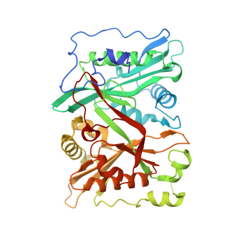
Human N-myristoyltransferase (NMT1) with Myristoyl-CoA and peptide bound


Experimental Data Snapshot

  • Method: X-RAY DIFFRACTION
  • Resolution: 2.10 Å
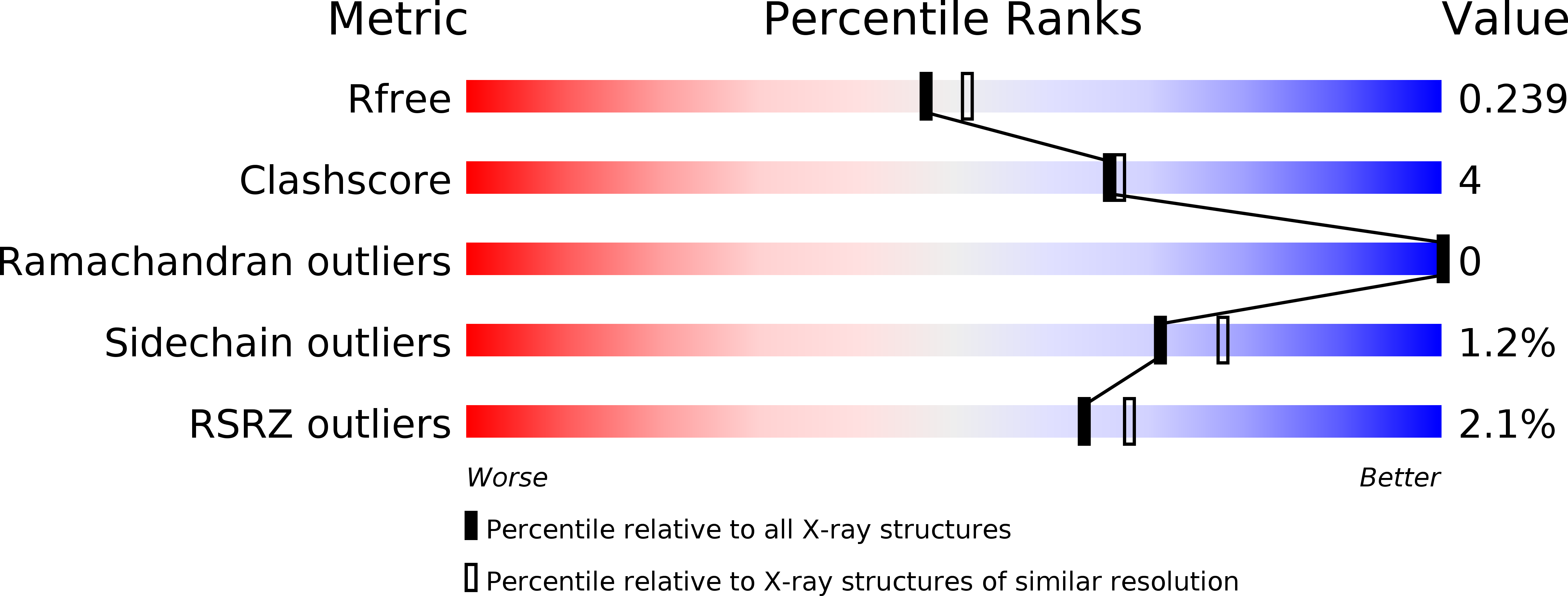
  • R-Value Free: 0.239 
  • R-Value Work: 0.184 
  • R-Value Observed: 0.187 

Starting Model: experimental
View more details

wwPDB Validation   3D Report Full Report


Ligand Structure Quality Assessment 


This is version 2.1 of the entry. See complete history


Literature

High-resolution snapshots of human N-myristoyltransferase in action illuminate a mechanism promoting N-terminal Lys and Gly myristoylation.

Dian, C.Perez-Dorado, I.Riviere, F.Asensio, T.Legrand, P.Ritzefeld, M.Shen, M.Cota, E.Meinnel, T.Tate, E.W.Giglione, C.

(2020) Nat Commun 11: 1132-1132

  • DOI: https://doi.org/10.1038/s41467-020-14847-3
  • Primary Citation of Related Structures:  
    6EHJ, 6QRM, 6SJZ, 6SK2, 6SK3, 6SK8, 6SKJ

  • PubMed Abstract: 

    The promising drug target N-myristoyltransferase (NMT) catalyses an essential protein modification thought to occur exclusively at N-terminal glycines (Gly). Here, we present high-resolution human NMT1 structures co-crystallised with reactive cognate lipid and peptide substrates, revealing high-resolution snapshots of the entire catalytic mechanism from the initial to final reaction states. Structural comparisons, together with biochemical analysis, provide unforeseen details about how NMT1 reaches a catalytically competent conformation in which the reactive groups are brought into close proximity to enable catalysis. We demonstrate that this mechanism further supports efficient and unprecedented myristoylation of an N-terminal lysine side chain, providing evidence that NMT acts both as N-terminal-lysine and glycine myristoyltransferase.


  • Organizational Affiliation

    Université Paris-Saclay, CEA, CNRS, Institute for Integrative Biology of the Cell (I2BC), Gif-sur-Yvette, 91198, France.


Macromolecules
Find similar proteins by:  (by identity cutoff)  |  3D Structure
Entity ID: 1
MoleculeChains Sequence LengthOrganismDetailsImage
Glycylpeptide N-tetradecanoyltransferase 1
A, B
391Homo sapiensMutation(s): 0 
Gene Names: NMT1NMT
EC: 2.3.1.97 (PDB Primary Data), 2.3.1 (UniProt)
UniProt & NIH Common Fund Data Resources
Find proteins for P30419 (Homo sapiens)
Explore P30419 
Go to UniProtKB:  P30419
PHAROS:  P30419
GTEx:  ENSG00000136448 
Entity Groups  
Sequence Clusters30% Identity50% Identity70% Identity90% Identity95% Identity100% Identity
UniProt GroupP30419
Sequence Annotations
Expand
  • Reference Sequence
Small Molecules
Ligands 9 Unique
IDChains Name / Formula / InChI Key2D Diagram3D Interactions
MYA (Subject of Investigation/LOI)
Query on MYA

Download Ideal Coordinates CCD File 
E [auth A]TETRADECANOYL-COA
C35 H62 N7 O17 P3 S
DUAFKXOFBZQTQE-QSGBVPJFSA-N
COA
Query on COA

Download Ideal Coordinates CCD File 
O [auth A],
R [auth B]
COENZYME A
C21 H36 N7 O16 P3 S
RGJOEKWQDUBAIZ-IBOSZNHHSA-N
MYR
Query on MYR

Download Ideal Coordinates CCD File 
N [auth A],
S [auth B]
MYRISTIC ACID
C14 H28 O2
TUNFSRHWOTWDNC-UHFFFAOYSA-N
LYS
Query on LYS

Download Ideal Coordinates CCD File 
AA [auth B]
I [auth A]
K [auth A]
M [auth A]
W [auth B]
AA [auth B],
I [auth A],
K [auth A],
M [auth A],
W [auth B],
Y [auth B]
LYSINE
C6 H15 N2 O2
KDXKERNSBIXSRK-YFKPBYRVSA-O
ASN
Query on ASN

Download Ideal Coordinates CCD File 
H [auth A],
V [auth B]
ASPARAGINE
C4 H8 N2 O3
DCXYFEDJOCDNAF-REOHCLBHSA-N
PRO
Query on PRO

Download Ideal Coordinates CCD File 
L [auth A],
Z [auth B]
PROLINE
C5 H9 N O2
ONIBWKKTOPOVIA-BYPYZUCNSA-N
SER
Query on SER

Download Ideal Coordinates CCD File 
G [auth A],
J [auth A],
U [auth B],
X [auth B]
SERINE
C3 H7 N O3
MTCFGRXMJLQNBG-REOHCLBHSA-N
GOL
Query on GOL

Download Ideal Coordinates CCD File 
C [auth A],
D [auth A],
P [auth B],
Q [auth B]
GLYCEROL
C3 H8 O3
PEDCQBHIVMGVHV-UHFFFAOYSA-N
GLY
Query on GLY

Download Ideal Coordinates CCD File 
F [auth A],
T [auth B]
GLYCINE
C2 H5 N O2
DHMQDGOQFOQNFH-UHFFFAOYSA-N
Experimental Data & Validation

Experimental Data

  • Method: X-RAY DIFFRACTION
  • Resolution: 2.10 Å
  • R-Value Free: 0.239 
  • R-Value Work: 0.184 
  • R-Value Observed: 0.187 
  • Space Group: P 21 21 2
Unit Cell:
Length ( Å )Angle ( ˚ )
a = 80.25α = 90
b = 177.54β = 90
c = 58.14γ = 90
Software Package:
Software NamePurpose
PHENIXrefinement
XDSdata reduction
Aimlessdata scaling
PHASERphasing

Structure Validation

View Full Validation Report


Ligand Structure Quality Assessment 


Entry History 

Deposition Data

Revision History  (Full details and data files)

  • Version 1.0: 2019-03-27
    Type: Initial release
  • Version 1.1: 2019-07-10
    Changes: Data collection
  • Version 2.0: 2020-04-22
    Type: Coordinate replacement
    Reason: Ligand geometry
    Changes: Advisory, Atomic model, Author supporting evidence, Data collection, Database references, Derived calculations, Non-polymer description, Polymer sequence, Refinement description, Source and taxonomy, Structure summary
  • Version 2.1: 2024-01-17
    Changes: Data collection, Database references, Refinement description