6OVQ | pdb_00006ovq

Crystal structure of mithramycin 3-side chain keto-reductase MtmW


Experimental Data Snapshot

  • Method: X-RAY DIFFRACTION
  • Resolution: 1.80 Å
  • R-Value Free: 
    0.213 (Depositor), 0.224 (DCC) 
  • R-Value Work: 
    0.184 (Depositor), 0.194 (DCC) 
  • R-Value Observed: 
    0.186 (Depositor) 

Starting Model: experimental
View more details

wwPDB Validation 3D Report Full Report

Validation slider image for 6OVQ

This is version 1.2 of the entry. See complete history

Literature

Discovery of a Cryptic Intermediate in Late Steps of Mithramycin Biosynthesis.

Wheeler, R.Yu, X.Hou, C.Mitra, P.Chen, J.M.Herkules, F.Ivanov, D.N.Tsodikov, O.V.Rohr, J.

(2020) Angew Chem Int Ed Engl 59: 826-832

  • DOI: https://doi.org/10.1002/anie.201910241
  • Primary Citation Related Structures: 
    6OVQ, 6OVX, 6OW0

  • PubMed Abstract: 

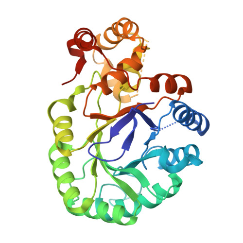
    MtmOIV and MtmW catalyze the final two reactions in the mithramycin (MTM) biosynthetic pathway, the Baeyer-Villiger opening of the fourth ring of premithramycin B (PMB), creating the C3 pentyl side chain, strictly followed by reduction of the distal keto group on the new side chain. Unexpectedly this results in a C2 stereoisomer of mithramycin, iso-mithramycin (iso-MTM). Iso-MTM undergoes a non-enzymatic isomerization to MTM catalyzed by Mg 2+ ions. Crystal structures of MtmW and its complexes with co-substrate NADPH and PEG, suggest a catalytic mechanism of MtmW. The structures also show that a tetrameric assembly of this enzyme strikingly resembles the ring-shaped β subunit of a vertebrate ion channel. We show that MtmW and MtmOIV form a complex in the presence of PMB and NADPH, presumably to hand over the unstable MtmOIV product to MtmW, yielding iso-MTM, as a potential self-resistance mechanism against MTM toxicity.


  • Organizational Affiliation
    • Department of Pharmaceutical Sciences, College of Pharmacy, University of Kentucky, 789 South Limestone Street, Lexington, KY, 40536-0596, USA.

Macromolecule Content 

  • Total Structure Weight: 72.86 kDa 
  • Atom Count: 4,845 
  • Modeled Residue Count: 586 
  • Deposited Residue Count: 666 
  • Unique protein chains: 1

Macromolecules

Find similar proteins by:|  3D Structure
Entity ID: 1
MoleculeChains  Sequence LengthOrganismDetailsImage
Putative Side chain reductase
A, B
333Streptomyces argillaceusMutation(s): 0 
Gene Names: mtmW
UniProt
Find proteins for Q194Q1 (Streptomyces argillaceus)
Explore Q194Q1 
Go to UniProtKB:  Q194Q1
Entity Groups
Sequence Clusters30% Identity50% Identity70% Identity90% Identity95% Identity100% Identity
UniProt GroupQ194Q1
Sequence Annotations
Expand
Reference Sequence

Experimental Data & Validation

Experimental Data

  • Method: X-RAY DIFFRACTION
  • Resolution: 1.80 Å
  • R-Value Free:  0.213 (Depositor), 0.224 (DCC) 
  • R-Value Work:  0.184 (Depositor), 0.194 (DCC) 
  • R-Value Observed: 0.186 (Depositor) 
Space Group: I 4
Unit Cell:
Length ( Å )Angle ( ˚ )
a = 131.105α = 90
b = 131.105β = 90
c = 79.769γ = 90
Software Package:
Software NamePurpose
REFMACrefinement
HKL-2000data reduction
HKL-2000data scaling
PHASERphasing

Structure Validation

View Full Validation Report


Entry History 

& Funding Information

Deposition Data


Funding OrganizationLocationGrant Number
National Institutes of Health/National Institute of General Medical Sciences (NIH/NIGMS)United StatesR01GM105977-01A1

Revision History  (Full details and data files)

  • Version 1.0: 2019-11-27
    Type: Initial release
  • Version 1.1: 2020-01-15
    Changes: Database references
  • Version 1.2: 2023-10-11
    Changes: Data collection, Database references, Refinement description