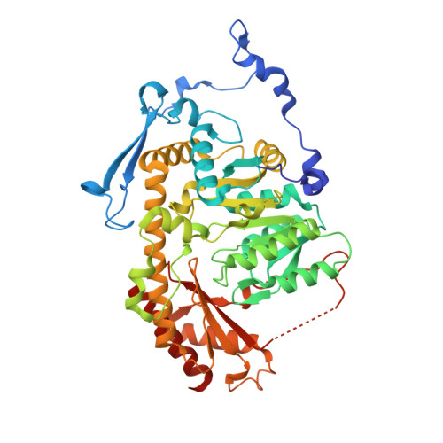
6HNU

Crystal structure of the aminotransferase Aro8 from C. Albicans with ligands


Experimental Data Snapshot

  • Method: X-RAY DIFFRACTION
  • Resolution: 1.80 Å
  • R-Value Free: 0.219 
  • R-Value Work: 0.174 
  • R-Value Observed: 0.176 

Starting Model: experimental
View more details

wwPDB Validation   3D Report Full Report


Ligand Structure Quality Assessment 


This is version 1.2 of the entry. See complete history


Literature

Crystal structures of aminotransferases Aro8 and Aro9 from Candida albicans and structural insights into their properties.

Kiliszek, A.Rypniewski, W.Rzad, K.Milewski, S.Gabriel, I.

(2019) J Struct Biol 205: 26-33

  • DOI: https://doi.org/10.1016/j.jsb.2019.02.001
  • Primary Citation of Related Structures:  
    6HNB, 6HND, 6HNU, 6HNV

  • PubMed Abstract: 

    Aminotransferases catalyze reversibly the transamination reaction by a ping-pong bi-bi mechanism with pyridoxal 5'-phosphate (PLP) as a cofactor. Various aminotransferases acting on a range of substrates have been reported. Aromatic transaminases are able to catalyze the transamination reaction with both aromatic and acidic substrates. Two aminotransferases from C. albicans, Aro8p and Aro9p, have been identified recently, exhibiting different catalytic properties. To elucidate the multiple substrate recognition of the two enzymes we determined the crystal structures of an unliganded CaAro8p, a complex of CaAro8p with the PLP cofactor bound to a substrate, forming an external aldimine, CaAro9p with PLP in the form of internal aldimine, and CaAro9p with a mixture of ligands that have been interpreted as results of the enzymatic reaction. The crystal structures of both enzymes contains in the asymmetric unit a biologically relevant dimer of 55 kDa for CaAro8 and 59 kDa for CaAro9p protein subunits. The ability of the enzymes to process multiple substrates could be related to a feature of their architecture in which the active site resides on one subunit while the substrate-binding site is formed by a long loop extending from the other subunit of the dimeric molecule. The separation of the two functions to different chemical entities could facilitate the evolution of the substrate-binding part and allow it to be flexible without destabilizing the conservative catalytic mechanism.


  • Organizational Affiliation

    Institute of Bioorganic Chemistry, Polish Academy of Sciences, Noskowskiego 12-14, 61-704 Poznań, Poland.


Macromolecules
Find similar proteins by:  (by identity cutoff)  |  3D Structure
Entity ID: 1
MoleculeChains Sequence LengthOrganismDetailsImage
Aromatic amino acid aminotransferase I
A, B
491Candida albicans WO-1Mutation(s): 0 
Gene Names: CAWG_03814
UniProt
Find proteins for C4YJ02 (Candida albicans (strain WO-1))
Explore C4YJ02 
Go to UniProtKB:  C4YJ02
Entity Groups  
Sequence Clusters30% Identity50% Identity70% Identity90% Identity95% Identity100% Identity
UniProt GroupC4YJ02
Sequence Annotations
Expand
  • Reference Sequence
Small Molecules
Ligands 5 Unique
IDChains Name / Formula / InChI Key2D Diagram3D Interactions
PLP
Query on PLP

Download Ideal Coordinates CCD File 
D [auth A],
I [auth B]
PYRIDOXAL-5'-PHOSPHATE
C8 H10 N O6 P
NGVDGCNFYWLIFO-UHFFFAOYSA-N
BTB
Query on BTB

Download Ideal Coordinates CCD File 
C [auth A],
H [auth B]
2-[BIS-(2-HYDROXY-ETHYL)-AMINO]-2-HYDROXYMETHYL-PROPANE-1,3-DIOL
C8 H19 N O5
OWMVSZAMULFTJU-UHFFFAOYSA-N
PHE
Query on PHE

Download Ideal Coordinates CCD File 
E [auth A],
J [auth B]
PHENYLALANINE
C9 H11 N O2
COLNVLDHVKWLRT-QMMMGPOBSA-N
TRS
Query on TRS

Download Ideal Coordinates CCD File 
K [auth B]2-AMINO-2-HYDROXYMETHYL-PROPANE-1,3-DIOL
C4 H12 N O3
LENZDBCJOHFCAS-UHFFFAOYSA-O
CL
Query on CL

Download Ideal Coordinates CCD File 
F [auth A],
G [auth A],
L [auth B],
M [auth B]
CHLORIDE ION
Cl
VEXZGXHMUGYJMC-UHFFFAOYSA-M
Experimental Data & Validation

Experimental Data

  • Method: X-RAY DIFFRACTION
  • Resolution: 1.80 Å
  • R-Value Free: 0.219 
  • R-Value Work: 0.174 
  • R-Value Observed: 0.176 
  • Space Group: P 21 21 21
Unit Cell:
Length ( Å )Angle ( ˚ )
a = 69.785α = 90
b = 102.09β = 90
c = 146.883γ = 90
Software Package:
Software NamePurpose
REFMACrefinement
XDSdata reduction
XDSdata scaling
PHASERphasing

Structure Validation

View Full Validation Report


Ligand Structure Quality Assessment 


Entry History & Funding Information

Deposition Data


Funding OrganizationLocationGrant Number
Poland2015/17/B/NZ6/04248

Revision History  (Full details and data files)

  • Version 1.0: 2019-02-20
    Type: Initial release
  • Version 1.1: 2019-03-27
    Changes: Data collection, Database references
  • Version 1.2: 2024-01-24
    Changes: Data collection, Database references, Refinement description