7KTK | pdb_00007ktk

DNA Polymerase Mu (K438D), 8-oxodGTP:Ct Ground State Ternary Complex, 50 mM Mg2+ (90min)


Experimental Data Snapshot

  • Method: X-RAY DIFFRACTION
  • Resolution: 1.42 Å
  • R-Value Free: 
    0.168 (Depositor), 0.178 (DCC) 
  • R-Value Work: 
    0.143 (Depositor), 0.154 (DCC) 
  • R-Value Observed: 
    0.144 (Depositor) 

Starting Model: experimental
View more details

wwPDB Validation 3D Report Full Report

Validation slider image for 7KTK

Ligand Structure Quality Assessment 


This is version 1.1 of the entry. See complete history

Literature

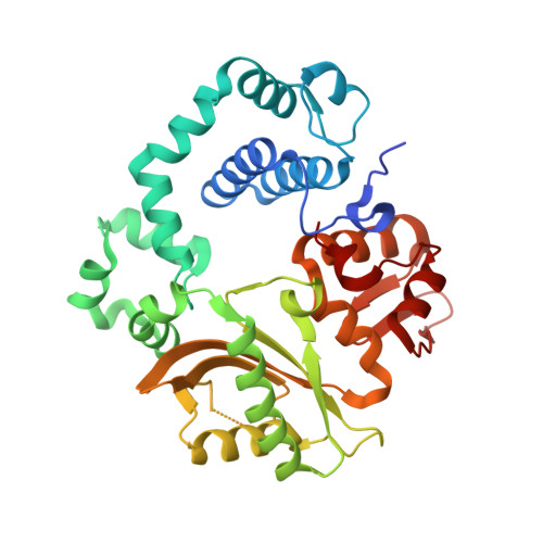
Watching a double strand break repair polymerase insert a pro-mutagenic oxidized nucleotide.

Jamsen, J.A.Sassa, A.Shock, D.D.Beard, W.A.Wilson, S.H.

(2021) Nat Commun 12: 2059-2059

  • DOI: https://doi.org/10.1038/s41467-021-21354-6
  • Primary Citation Related Structures: 
    7KSS, 7KST, 7KSU, 7KSV, 7KSW, 7KSX, 7KSY, 7KSZ, 7KT0, 7KT1, 7KT2, 7KT3, 7KT4, 7KT5, 7KT6, 7KT7, 7KT8, 7KT9, 7KTA, 7KTB, 7KTC, 7KTD, 7KTE, 7KTF, 7KTG, 7KTH, 7KTI, 7KTJ, 7KTK, 7KTL, 7KTM, 7KTN

  • PubMed Abstract: 

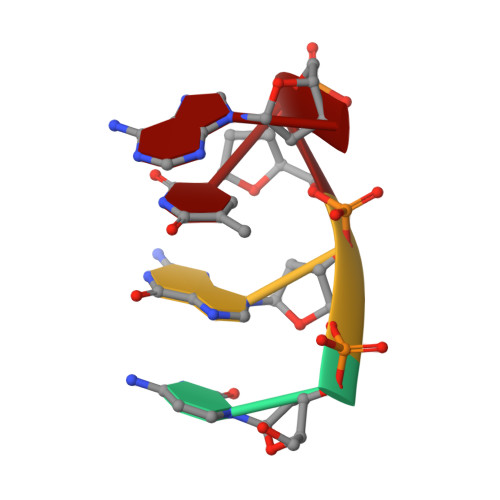
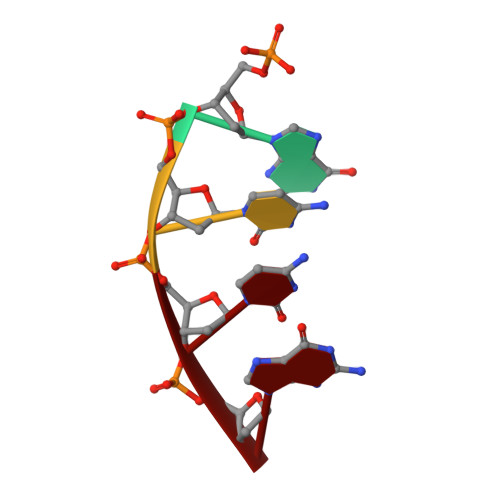
    Oxidized dGTP (8-oxo-7,8-dihydro-2´-deoxyguanosine triphosphate, 8-oxodGTP) insertion by DNA polymerases strongly promotes cancer and human disease. How DNA polymerases discriminate against oxidized and undamaged nucleotides, especially in error-prone double strand break (DSB) repair, is poorly understood. High-resolution time-lapse X-ray crystallography snapshots of DSB repair polymerase μ undergoing DNA synthesis reveal that a third active site metal promotes insertion of oxidized and undamaged dGTP in the canonical anti-conformation opposite template cytosine. The product metal bridged O8 with product oxygens, and was not observed in the syn-conformation opposite template adenine (A t ). Rotation of A t into the syn-conformation enabled undamaged dGTP misinsertion. Exploiting metal and substrate dynamics in a rigid active site allows 8-oxodGTP to circumvent polymerase fidelity safeguards to promote pro-mutagenic double strand break repair.


  • Organizational Affiliation
    • Genome Integrity and Structural Biology Laboratory, National Institute of Environmental Health Sciences, National Institutes of Health, Research Triangle Park, NC, 27709, USA.

Macromolecule Content 

  • Total Structure Weight: 46.66 kDa 
  • Atom Count: 3,544 
  • Modeled Residue Count: 346 
  • Deposited Residue Count: 373 
  • Unique protein chains: 1
  • Unique nucleic acid chains: 3

Macromolecules


Find similar proteins by:|  3D Structure
Entity ID: 1
MoleculeChains  Sequence LengthOrganismDetailsImage
DNA-directed DNA/RNA polymerase mu356Homo sapiensMutation(s): 1 
Gene Names: POLMpolmu
EC: 2.7.7.7
UniProt & NIH Common Fund Data Resources
Find proteins for Q9NP87 (Homo sapiens)
Explore Q9NP87 
Go to UniProtKB:  Q9NP87
PHAROS:  Q9NP87
GTEx:  ENSG00000122678 
Entity Groups
Sequence Clusters30% Identity50% Identity70% Identity90% Identity95% Identity100% Identity
UniProt GroupQ9NP87
Sequence Annotations
Expand
Reference Sequence
Find similar nucleic acids by:  Sequence
Entity ID: 2
MoleculeChains LengthOrganismImage
DNA (5'-D(*CP*GP*GP*CP*CP*TP*AP*CP*G)-3')B [auth T]9synthetic construct
Sequence Annotations
Expand
Reference Sequence
Find similar nucleic acids by:  Sequence
Entity ID: 3
MoleculeChains LengthOrganismImage
DNA (5'-D(*CP*GP*TP*A)-3')C [auth P]4synthetic construct
Sequence Annotations
Expand
Reference Sequence
Find similar nucleic acids by:  Sequence
Entity ID: 4
MoleculeChains LengthOrganismImage
DNA (5'-D(P*GP*CP*CP*G)-3')4synthetic construct
Sequence Annotations
Expand
Reference Sequence

Small Molecules

Ligands 5 Unique
IDChains Name / Formula / InChI Key2D Diagram3D Interactions
8DG
(Subject of Investigation/LOI)

Query on 8DG


Download:Ideal Coordinates CCD File
E [auth A]8-OXO-2'-DEOXYGUANOSINE-5'-TRIPHOSPHATE
C10 H16 N5 O14 P3
BUZOGVVQWCXXDP-VPENINKCSA-N
EPE

Query on EPE


Download:Ideal Coordinates CCD File
I [auth A]4-(2-HYDROXYETHYL)-1-PIPERAZINE ETHANESULFONIC ACID
C8 H18 N2 O4 S
JKMHFZQWWAIEOD-UHFFFAOYSA-N
EDO

Query on EDO


Download:Ideal Coordinates CCD File
J [auth A]
K [auth A]
L [auth A]
M [auth A]
N [auth A]
J [auth A],
K [auth A],
L [auth A],
M [auth A],
N [auth A],
O [auth A],
P [auth A],
Q [auth A],
R [auth A],
S [auth A],
T [auth D]
1,2-ETHANEDIOL
C2 H6 O2
LYCAIKOWRPUZTN-UHFFFAOYSA-N
MG
(Subject of Investigation/LOI)

Query on MG


Download:Ideal Coordinates CCD File
F [auth A],
G [auth A]
MAGNESIUM ION
Mg
JLVVSXFLKOJNIY-UHFFFAOYSA-N
NA

Query on NA


Download:Ideal Coordinates CCD File
H [auth A]SODIUM ION
Na
FKNQFGJONOIPTF-UHFFFAOYSA-N

Experimental Data & Validation

Experimental Data

  • Method: X-RAY DIFFRACTION
  • Resolution: 1.42 Å
  • R-Value Free:  0.168 (Depositor), 0.178 (DCC) 
  • R-Value Work:  0.143 (Depositor), 0.154 (DCC) 
  • R-Value Observed: 0.144 (Depositor) 
Space Group: P 21 21 21
Unit Cell:
Length ( Å )Angle ( ˚ )
a = 60.109α = 90
b = 61.775β = 90
c = 118.076γ = 90
Software Package:
Software NamePurpose
HKL-2000data reduction
HKL-2000data scaling
PHENIXrefinement
PDB_EXTRACTdata extraction
PHENIXphasing

Structure Validation

View Full Validation Report


Ligand Structure Quality Assessment 


Entry History 

& Funding Information

Deposition Data


Funding OrganizationLocationGrant Number
National Institutes of Health/National Institute of Environmental Health Sciences (NIH/NIEHS)United StatesZ01-ES050158
National Institutes of Health/National Institute of Environmental Health Sciences (NIH/NIEHS)United StatesZ01-ES050161
Japan Society for the Promotion of Science (JSPS)Japan16K16195

Revision History  (Full details and data files)

  • Version 1.0: 2022-05-11
    Type: Initial release
  • Version 1.1: 2023-10-18
    Changes: Data collection, Refinement description