7YCM

TR-SFX MmCPDII-DNA complex: 100 ps snapshot. Includes 100ps, dark, and extrapolated structure factors


Experimental Data Snapshot

  • Method: X-RAY DIFFRACTION
  • Resolution: 2.00 Å
  • R-Value Free: 0.212 
  • R-Value Work: 0.193 
  • R-Value Observed: 0.194 

wwPDB Validation   3D Report Full Report


Ligand Structure Quality Assessment 


This is version 1.2 of the entry. See complete history


Literature

Visualizing the DNA repair process by a photolyase at atomic resolution.

Maestre-Reyna, M.Wang, P.H.Nango, E.Hosokawa, Y.Saft, M.Furrer, A.Yang, C.H.Gusti Ngurah Putu, E.P.Wu, W.J.Emmerich, H.J.Caramello, N.Franz-Badur, S.Yang, C.Engilberge, S.Wranik, M.Glover, H.L.Weinert, T.Wu, H.Y.Lee, C.C.Huang, W.C.Huang, K.F.Chang, Y.K.Liao, J.H.Weng, J.H.Gad, W.Chang, C.W.Pang, A.H.Yang, K.C.Lin, W.T.Chang, Y.C.Gashi, D.Beale, E.Ozerov, D.Nass, K.Knopp, G.Johnson, P.J.M.Cirelli, C.Milne, C.Bacellar, C.Sugahara, M.Owada, S.Joti, Y.Yamashita, A.Tanaka, R.Tanaka, T.Luo, F.Tono, K.Zarzycka, W.Muller, P.Alahmad, M.A.Bezold, F.Fuchs, V.Gnau, P.Kiontke, S.Korf, L.Reithofer, V.Rosner, C.J.Seiler, E.M.Watad, M.Werel, L.Spadaccini, R.Yamamoto, J.Iwata, S.Zhong, D.Standfuss, J.Royant, A.Bessho, Y.Essen, L.O.Tsai, M.D.

(2023) Science 382: eadd7795-eadd7795

  • DOI: https://doi.org/10.1126/science.add7795
  • Primary Citation of Related Structures:  
    7YC7, 7YCM, 7YCP, 7YCR, 7YD6, 7YD7, 7YD8, 7YDZ, 7YE0, 7YEB, 7YEC, 7YEE, 7YEI, 7YEJ, 7YEK, 7YEL, 7YEM, 8KCM

  • PubMed Abstract: 

    Photolyases, a ubiquitous class of flavoproteins, use blue light to repair DNA photolesions. In this work, we determined the structural mechanism of the photolyase-catalyzed repair of a cyclobutane pyrimidine dimer (CPD) lesion using time-resolved serial femtosecond crystallography (TR-SFX). We obtained 18 snapshots that show time-dependent changes in four reaction loci. We used these results to create a movie that depicts the repair of CPD lesions in the picosecond-to-nanosecond range, followed by the recovery of the enzymatic moieties involved in catalysis, completing the formation of the fully reduced enzyme-product complex at 500 nanoseconds. Finally, back-flip intermediates of the thymine bases to reanneal the DNA were captured at 25 to 200 microseconds. Our data cover the complete molecular mechanism of a photolyase and, importantly, its chemistry and enzymatic catalysis at work across a wide timescale and at atomic resolution.


  • Organizational Affiliation

    Institute of Biological Chemistry, Academia Sinica, 128 Academia Rd. Sec. 2, Nankang, Taipei 115, Taiwan.


Macromolecules

Find similar proteins by:  (by identity cutoff)  |  3D Structure
Entity ID: 1
MoleculeChains Sequence LengthOrganismDetailsImage
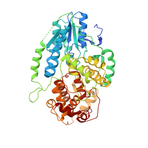
Deoxyribodipyrimidine photolyase
A, B
482Methanosarcina mazei Go1Mutation(s): 1 
EC: 4.1.99.3
UniProt
Find proteins for Q8PYK9 (Methanosarcina mazei (strain ATCC BAA-159 / DSM 3647 / Goe1 / Go1 / JCM 11833 / OCM 88))
Explore Q8PYK9 
Go to UniProtKB:  Q8PYK9
Entity Groups  
Sequence Clusters30% Identity50% Identity70% Identity90% Identity95% Identity100% Identity
UniProt GroupQ8PYK9
Sequence Annotations
Expand
  • Reference Sequence

Find similar nucleic acids by:  Sequence   |   3D Structure  

Entity ID: 2
MoleculeChains LengthOrganismImage
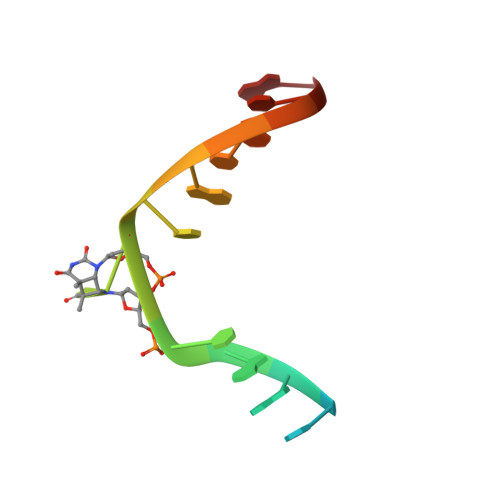
CPD photolesion containing DNA
C, E
14synthetic construct
Sequence Annotations
Expand
  • Reference Sequence

Find similar nucleic acids by:  Sequence   |   3D Structure  

Entity ID: 3
MoleculeChains LengthOrganismImage
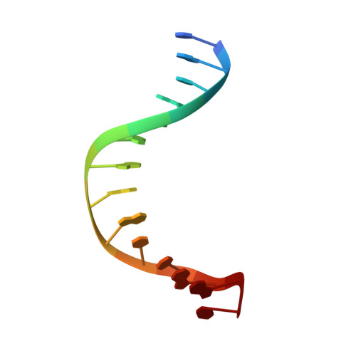
complementary oligonucleotide to the CPD containing DNA
D, F
14synthetic construct
Sequence Annotations
Expand
  • Reference Sequence
Experimental Data & Validation

Experimental Data

  • Method: X-RAY DIFFRACTION
  • Resolution: 2.00 Å
  • R-Value Free: 0.212 
  • R-Value Work: 0.193 
  • R-Value Observed: 0.194 
  • Space Group: P 21 21 21
Unit Cell:
Length ( Å )Angle ( ˚ )
a = 72.47α = 90
b = 115.93β = 90
c = 169.35γ = 90
Software Package:
Software NamePurpose
PHENIXrefinement
CrystFELdata reduction
pointlessdata scaling
REFMACphasing

Structure Validation

View Full Validation Report


Ligand Structure Quality Assessment 


Entry History & Funding Information

Deposition Data


Funding OrganizationLocationGrant Number
Ministry of Science and Technology (MoST, Taiwan)TaiwanAS-KPQ-105-TPP
Ministry of Science and Technology (MoST, Taiwan)TaiwanAS-KPQ-109-TPP2
Japan Society for the Promotion of Science (JSPS)Japan16K01942

Revision History  (Full details and data files)

  • Version 1.0: 2023-11-22
    Type: Initial release
  • Version 1.1: 2023-12-13
    Changes: Database references
  • Version 1.2: 2024-03-13
    Changes: Source and taxonomy