7ZKI

Cryo-EM structure of aIF1A:aIF5B:Met-tRNAiMet complex from a Pyrococcus abyssi 30S initiation complex


Experimental Data Snapshot

  • Method: ELECTRON MICROSCOPY
  • Resolution: 3.60 Å
  • Aggregation State: PARTICLE 
  • Reconstruction Method: SINGLE PARTICLE 

wwPDB Validation   3D Report Full Report

Currently 7ZKI does not have a validation slider image.


This is version 1.1 of the entry. See complete history


Literature

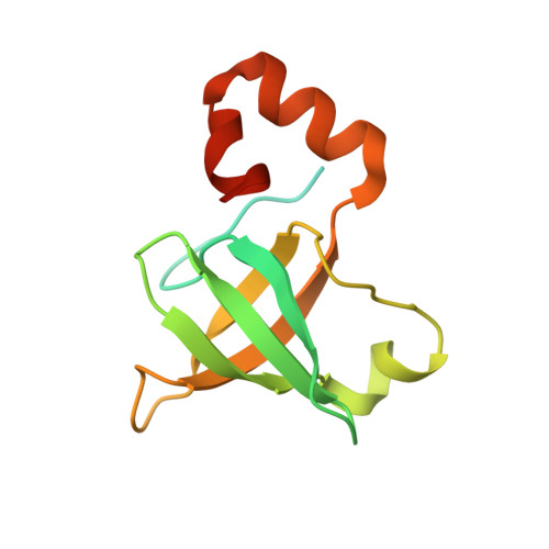
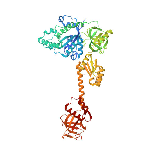
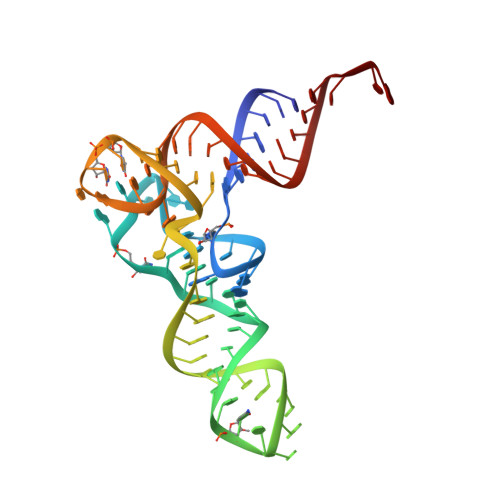
Role of aIF5B in archaeal translation initiation.

Kazan, R.Bourgeois, G.Lazennec-Schurdevin, C.Larquet, E.Mechulam, Y.Coureux, P.D.Schmitt, E.

(2022) Nucleic Acids Res 50: 6532-6548

  • DOI: https://doi.org/10.1093/nar/gkac490
  • Primary Citation of Related Structures:  
    7YYP, 7YZN, 7ZAG, 7ZAH, 7ZAI, 7ZHG, 7ZKI

  • PubMed Abstract: 

    In eukaryotes and in archaea late steps of translation initiation involve the two initiation factors e/aIF5B and e/aIF1A. In eukaryotes, the role of eIF5B in ribosomal subunit joining is established and structural data showing eIF5B bound to the full ribosome were obtained. To achieve its function, eIF5B collaborates with eIF1A. However, structural data illustrating how these two factors interact on the small ribosomal subunit have long been awaited. The role of the archaeal counterparts, aIF5B and aIF1A, remains to be extensively addressed. Here, we study the late steps of Pyrococcus abyssi translation initiation. Using in vitro reconstituted initiation complexes and light scattering, we show that aIF5B bound to GTP accelerates subunit joining without the need for GTP hydrolysis. We report the crystallographic structures of aIF5B bound to GDP and GTP and analyze domain movements associated to these two nucleotide states. Finally, we present the cryo-EM structure of an initiation complex containing 30S bound to mRNA, Met-tRNAiMet, aIF5B and aIF1A at 2.7 Å resolution. Structural data shows how archaeal 5B and 1A factors cooperate to induce a conformation of the initiator tRNA favorable to subunit joining. Archaeal and eukaryotic features of late steps of translation initiation are discussed.


  • Organizational Affiliation

    Laboratoire de Biologie Structurale de la Cellule, BIOC, Ecole polytechnique, CNRS, Institut Polytechnique de Paris, 91128 Palaiseau cedex, France.


Macromolecules

Find similar proteins by:  (by identity cutoff)  |  3D Structure
Entity ID: 2
MoleculeChains Sequence LengthOrganismDetailsImage
Translation initiation factor 1AB [auth 6]134Pyrococcus abyssi GE5Mutation(s): 0 
Gene Names: eIF1Aaif1APYRAB05910PAB2441
UniProt
Find proteins for Q9V138 (Pyrococcus abyssi (strain GE5 / Orsay))
Explore Q9V138 
Go to UniProtKB:  Q9V138
Entity Groups  
Sequence Clusters30% Identity50% Identity70% Identity90% Identity95% Identity100% Identity
UniProt GroupQ9V138
Sequence Annotations
Expand
  • Reference Sequence
Find similar proteins by:  (by identity cutoff)  |  3D Structure
Entity ID: 3
MoleculeChains Sequence LengthOrganismDetailsImage
Translation initiation factor 5BC [auth 7]619Pyrococcus abyssi GE5Mutation(s): 0 
Gene Names: WBA_LOCUS12468infBPYRAB11390PAB0755
UniProt
Find proteins for Q9UZK7 (Pyrococcus abyssi (strain GE5 / Orsay))
Explore Q9UZK7 
Go to UniProtKB:  Q9UZK7
Entity Groups  
Sequence Clusters30% Identity50% Identity70% Identity90% Identity95% Identity100% Identity
UniProt GroupQ9UZK7
Sequence Annotations
Expand
  • Reference Sequence
Find similar nucleic acids by:  (by identity cutoff)  |  3D Structure
Entity ID: 1
MoleculeChains LengthOrganismImage
Met-tRNAiMetA [auth 4]77Escherichia coli
Sequence Annotations
Expand
  • Reference Sequence
Experimental Data & Validation

Experimental Data

  • Method: ELECTRON MICROSCOPY
  • Resolution: 3.60 Å
  • Aggregation State: PARTICLE 
  • Reconstruction Method: SINGLE PARTICLE 

Structure Validation

View Full Validation Report

Currently 7ZKI does not have a validation slider image.


Entry History & Funding Information

Deposition Data


Funding OrganizationLocationGrant Number
Agence Nationale de la Recherche (ANR)France--

Revision History  (Full details and data files)

  • Version 1.0: 2022-07-20
    Type: Initial release
  • Version 1.1: 2022-08-31
    Changes: Database references