8A3P

Structure of human Fy-5.


Experimental Data Snapshot

  • Method: X-RAY DIFFRACTION
  • Resolution: 2.70 Å
  • R-Value Free: 0.267 
  • R-Value Work: 0.220 
  • R-Value Observed: 0.225 

wwPDB Validation   3D Report Full Report


This is version 1.3 of the entry. See complete history


Literature

Structural basis of mRNA binding by the human FERRY Rab5 effector complex.

Quentin, D.Schuhmacher, J.S.Klink, B.U.Lauer, J.Shaikh, T.R.Huis In 't Veld, P.J.Welp, L.M.Urlaub, H.Zerial, M.Raunser, S.

(2023) Mol Cell 83: 1856-1871.e9

  • DOI: https://doi.org/10.1016/j.molcel.2023.05.009
  • Primary Citation of Related Structures:  
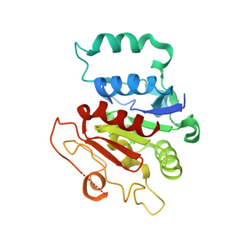
    7ND2, 8A3O, 8A3P

  • PubMed Abstract: 

    The pentameric FERRY Rab5 effector complex is a molecular link between mRNA and early endosomes in mRNA intracellular distribution. Here, we determine the cryo-EM structure of human FERRY. It reveals a unique clamp-like architecture that bears no resemblance to any known structure of Rab effectors. A combination of functional and mutational studies reveals that while the Fy-2 C-terminal coiled-coil acts as binding region for Fy-1/3 and Rab5, both coiled-coils and Fy-5 concur to bind mRNA. Mutations causing truncations of Fy-2 in patients with neurological disorders impair Rab5 binding or FERRY complex assembly. Thus, Fy-2 serves as a binding hub connecting all five complex subunits and mediating the binding to mRNA and early endosomes via Rab5. Our study provides mechanistic insights into long-distance mRNA transport and demonstrates that the particular architecture of FERRY is closely linked to a previously undescribed mode of RNA binding, involving coiled-coil domains.


  • Organizational Affiliation

    Department of Structural Biochemistry, Max Planck Institute of Molecular Physiology, 44227 Dortmund, Germany.


Macromolecules
Find similar proteins by:  (by identity cutoff)  |  3D Structure
Entity ID: 1
MoleculeChains Sequence LengthOrganismDetailsImage
Glutamine amidotransferase-like class 1 domain-containing protein 1220Homo sapiensMutation(s): 0 
Gene Names: GATD1PDDC1
UniProt & NIH Common Fund Data Resources
Find proteins for Q8NB37 (Homo sapiens)
Explore Q8NB37 
Go to UniProtKB:  Q8NB37
PHAROS:  Q8NB37
GTEx:  ENSG00000177225 
Entity Groups  
Sequence Clusters30% Identity50% Identity70% Identity90% Identity95% Identity100% Identity
UniProt GroupQ8NB37
Sequence Annotations
Expand
  • Reference Sequence
Experimental Data & Validation

Experimental Data

  • Method: X-RAY DIFFRACTION
  • Resolution: 2.70 Å
  • R-Value Free: 0.267 
  • R-Value Work: 0.220 
  • R-Value Observed: 0.225 
  • Space Group: C 2 2 21
Unit Cell:
Length ( Å )Angle ( ˚ )
a = 94.619α = 90
b = 99.573β = 90
c = 62.158γ = 90
Software Package:
Software NamePurpose
PHENIXrefinement
XDSdata reduction
SCALAdata scaling
PHASERphasing
PDB_EXTRACTdata extraction

Structure Validation

View Full Validation Report


Entry History & Funding Information

Deposition Data


Funding OrganizationLocationGrant Number
German Research Foundation (DFG)Germany112927078
Max Planck SocietyGermany--

Revision History  (Full details and data files)

  • Version 1.0: 2022-06-29
    Type: Initial release
  • Version 1.1: 2022-07-20
    Changes: Structure summary
  • Version 1.2: 2023-06-14
    Changes: Database references
  • Version 1.3: 2024-02-07
    Changes: Data collection, Refinement description