8PSV | pdb_00008psv

2.7 A cryo-EM structure of in vitro assembled type 1 pilus rod


Experimental Data Snapshot

  • Method: ELECTRON MICROSCOPY
  • Resolution: 2.70 Å
  • Aggregation State: FILAMENT 
  • Reconstruction Method: HELICAL 

Starting Model: experimental
View more details

wwPDB Validation 3D Report Full Report

Validation slider image for 8PSV

This is version 1.2 of the entry. See complete history

Literature

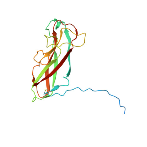
The assembly platform FimD is required to obtain the most stable quaternary structure of type 1 pili.

Zyla, D.S.Wiegand, T.Bachmann, P.Zdanowicz, R.Giese, C.Meier, B.H.Waksman, G.Hospenthal, M.K.Glockshuber, R.

(2024) Nat Commun 15: 3032-3032

  • DOI: https://doi.org/10.1038/s41467-024-47212-9
  • Primary Citation Related Structures: 
    6Y7S, 8PSV, 8PTU

  • PubMed Abstract: 

    Type 1 pili are important virulence factors of uropathogenic Escherichia coli that mediate bacterial attachment to epithelial cells in the urinary tract. The pilus rod is comprised of thousands of copies of the main structural subunit FimA and is assembled in vivo by the assembly platform FimD. Although type 1 pilus rods can self-assemble from FimA in vitro, this reaction is slower and produces structures with lower kinetic stability against denaturants compared to in vivo-assembled rods. Our study reveals that FimD-catalysed in vitro-assembled type 1 pilus rods attain a similar stability as pilus rods assembled in vivo. Employing structural, biophysical and biochemical analyses, we show that in vitro assembly reactions lacking FimD produce pilus rods with structural defects, reducing their stability against dissociation. Overall, our results indicate that FimD is not only required for the catalysis of pilus assembly, but also to control the assembly of the most stable quaternary structure.


  • Organizational Affiliation
    • Institute of Molecular Biology and Biophysics, ETH Zürich, Otto-Stern-Weg 5, 8093, Zürich, Switzerland.

Macromolecule Content 

  • Total Structure Weight: 108.73 kDa 
  • Atom Count: 6,642 
  • Modeled Residue Count: 948 
  • Deposited Residue Count: 1,092 
  • Unique protein chains: 1

Macromolecules

Find similar proteins by:|  3D Structure
Entity ID: 1
MoleculeChains  Sequence LengthOrganismDetailsImage
Type-1 fimbrial protein, A chain
A, B, C, D, E
A, B, C, D, E, F
182Escherichia coliMutation(s): 0 
Gene Names: fimApilAb4314JW4277
UniProt
Find proteins for P04128 (Escherichia coli (strain K12))
Explore P04128 
Go to UniProtKB:  P04128
Entity Groups
Sequence Clusters30% Identity50% Identity70% Identity90% Identity95% Identity100% Identity
UniProt GroupP04128
Sequence Annotations
Expand
Reference Sequence

Experimental Data & Validation

Experimental Data

  • Method: ELECTRON MICROSCOPY
  • Resolution: 2.70 Å
  • Aggregation State: FILAMENT 
  • Reconstruction Method: HELICAL 
EM Software:
TaskSoftware PackageVersion
RECONSTRUCTIONRELION3.08
MODEL REFINEMENTPHENIX

Structure Validation

View Full Validation Report


Entry History 

& Funding Information

Deposition Data


Funding OrganizationLocationGrant Number
Swiss National Science FoundationSwitzerland310030B_176403/1
Swiss National Science FoundationSwitzerland31003A_156304
Swiss National Science FoundationSwitzerland310030_201234
Medical Research Council (MRC, United Kingdom)United Kingdom018434

Revision History  (Full details and data files)

  • Version 1.0: 2024-04-10
    Type: Initial release
  • Version 1.1: 2024-04-17
    Changes: Database references
  • Version 1.2: 2024-11-13
    Changes: Data collection, Structure summary