Molecular principles of the assembly and construction of a carboxysome shell.
Wang, P., Li, J., Li, T., Li, K., Ng, P.C., Wang, S., Chriscoli, V., Basle, A., Marles-Wright, J., Zhang, Y.Z., Liu, L.N.(2024) Sci Adv 10: eadr4227-eadr4227
- PubMed: 39612341 Search on PubMedSearch on PubMed Central
- DOI: https://doi.org/10.1126/sciadv.adr4227
- Primary Citation Related Structures:
8YVC, 8YVD, 8YVE, 8YVF, 8YVI, 8YXU, 9F0H - PubMed Abstract:
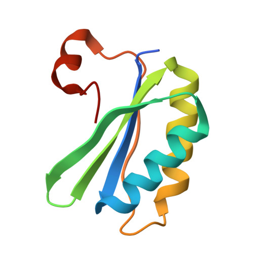
Intracellular compartmentalization enhances biological reactions, crucial for cellular function and survival. An example is the carboxysome, a bacterial microcompartment for CO 2 fixation. The carboxysome uses a polyhedral protein shell made of hexamers, pentamers, and trimers to encapsulate Rubisco, increasing CO 2 levels near Rubisco to enhance carboxylation. Despite their role in the global carbon cycle, the molecular mechanisms behind carboxysome shell assembly remain unclear. Here, we present a structural characterization of α-carboxysome shells generated from recombinant systems, which contain all shell proteins and the scaffolding protein CsoS2. Atomic-resolution cryo-electron microscopy of the shell assemblies, with a maximal size of 54 nm, unveil diverse assembly interfaces between shell proteins, detailed interactions of CsoS2 with shell proteins to drive shell assembly, and the formation of heterohexamers and heteropentamers by different shell protein paralogs, facilitating the assembly of larger empty shells. Our findings provide mechanistic insights into the construction principles of α-carboxysome shells and the role of CsoS2 in governing α-carboxysome assembly and functionality.
- MOE Key Laboratory of Evolution and Marine Biodiversity, Frontiers Science Center for Deep Ocean Multispheres and Earth System & College of Marine Life Sciences, Ocean University of China, Qingdao 266003, China.
Organizational Affiliation: