9B28

Cryo-EM structure of the mouse TRPM3 alpha 2 channel in complex with primidone


Experimental Data Snapshot

  • Method: ELECTRON MICROSCOPY
  • Resolution: 3.24 Å
  • Aggregation State: PARTICLE 
  • Reconstruction Method: SINGLE PARTICLE 

Starting Model: experimental
View more details

wwPDB Validation   3D Report Full Report


This is version 1.1 of the entry. See complete history


Literature

Molecular basis of neurosteroid and anticonvulsant regulation of TRPM3.

Yin, Y.Park, C.G.Feng, S.Guan, Z.Lee, H.J.Zhang, F.Sharma, K.Borgnia, M.J.Im, W.Lee, S.Y.

(2025) Nat Struct Mol Biol 

  • DOI: https://doi.org/10.1038/s41594-024-01463-8
  • Primary Citation of Related Structures:  
    9B28, 9B29, 9B2A

  • PubMed Abstract: 

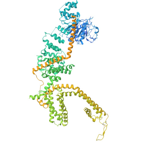
    Transient receptor potential channel subfamily M member 3 (TRPM3) is a Ca 2+ -permeable cation channel activated by the neurosteroid pregnenolone sulfate (PregS) or heat, serving as a nociceptor in the peripheral sensory system. Recent discoveries of autosomal dominant neurodevelopmental disorders caused by gain-of-function mutations in TRPM3 highlight its role in the central nervous system. Notably, the TRPM3 inhibitor primidone, an anticonvulsant, has proven effective in treating patients with TRPM3-linked neurological disorders and in mouse models of thermal nociception. However, our understanding of neurosteroids, inhibitors and disease mutations on TRPM3 is limited. Here we present cryogenic electron microscopy structures of the mouse TRPM3 in complex with cholesteryl hemisuccinate, primidone and PregS with the synthetic agonist CIM 0216. Our studies identify the binding sites for the neurosteroid, synthetic agonist and inhibitor and offer insights into their effects and disease mutations on TRPM3 gating, aiding future drug development.


  • Organizational Affiliation

    Department of Biochemistry, Duke University School of Medicine, Durham, NC, USA.


Macromolecules
Find similar proteins by:  (by identity cutoff)  |  3D Structure
Entity ID: 1
MoleculeChains Sequence LengthOrganismDetailsImage
Transient receptor potential cation channel, subfamily M, member 3A,
B [auth C],
C [auth B],
D
1,739Mus musculusMutation(s): 0 
Gene Names: Trpm3
UniProt & NIH Common Fund Data Resources
Find proteins for J9SQF3 (Mus musculus)
Explore J9SQF3 
Go to UniProtKB:  J9SQF3
IMPC:  MGI:2443101
Entity Groups  
Sequence Clusters30% Identity50% Identity70% Identity90% Identity95% Identity100% Identity
UniProt GroupJ9SQF3
Sequence Annotations
Expand
  • Reference Sequence
Small Molecules
Ligands 3 Unique
IDChains Name / Formula / InChI Key2D Diagram3D Interactions
6OU
Query on 6OU

Download Ideal Coordinates CCD File 
F [auth A],
J [auth C],
N [auth B],
R [auth D]
[(2~{R})-1-[2-azanylethoxy(oxidanyl)phosphoryl]oxy-3-hexadecanoyloxy-propan-2-yl] (~{Z})-octadec-9-enoate
C39 H76 N O8 P
FHQVHHIBKUMWTI-OTMQOFQLSA-N
Y01
Query on Y01

Download Ideal Coordinates CCD File 
E [auth A]
H [auth A]
I [auth C]
L [auth C]
M [auth B]
CHOLESTEROL HEMISUCCINATE
C31 H50 O4
WLNARFZDISHUGS-MIXBDBMTSA-N
A1AIA
Query on A1AIA

Download Ideal Coordinates CCD File 
G [auth A],
K [auth C],
O [auth B],
S [auth D]
primidone
C12 H14 N2 O2
DQMZLTXERSFNPB-UHFFFAOYSA-N
Experimental Data & Validation

Experimental Data

  • Method: ELECTRON MICROSCOPY
  • Resolution: 3.24 Å
  • Aggregation State: PARTICLE 
  • Reconstruction Method: SINGLE PARTICLE 
EM Software:
TaskSoftware PackageVersion
MODEL REFINEMENTPHENIX
RECONSTRUCTIONcryoSPARC

Structure Validation

View Full Validation Report


Entry History & Funding Information

Deposition Data


Funding OrganizationLocationGrant Number
National Institutes of Health/National Institute of Neurological Disorders and Stroke (NIH/NINDS)United States--

Revision History  (Full details and data files)

  • Version 1.0: 2025-01-22
    Type: Initial release
  • Version 1.1: 2025-01-29
    Changes: Data collection, Database references