9ZES | pdb_00009zes

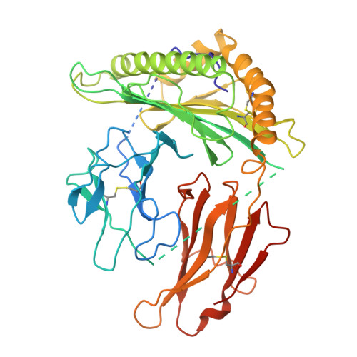
Germline-like TCR156 bound to single-chain poly-Ala-B2M-HLA-A*02


Experimental Data Snapshot

  • Method: ELECTRON MICROSCOPY
  • Resolution: 3.20 Å
  • Aggregation State: PARTICLE 
  • Reconstruction Method: SINGLE PARTICLE 

wwPDB Validation 3D Report Full Report

Validation slider image for 9ZES

This is version 1.0 of the entry. See complete history. 

Literature

MHC restriction is structurally encoded by the T cell receptor germline.

Wang, N., Borowska, M.T., Jude, K.M., Chen, X., Wang, C., Xiang, X., Liu, L.D., Ma, R., Motiani, A., Wilson, S.C., Liu, W., Garcia, K.C.

(2026) bioRxiv 

  • DOI: https://doi.org/10.64898/2026.09.24.754241
  • Primary Citation Related Structures: 
    11OE, 9ZEP, 9ZEQ, 9ZER, 9ZES, 9ZET, 9ZEU, 9ZEV

  • PubMed Abstract: 

    MHC restriction - the requirement for αβ T cell receptors (TCRs) to recognize Major Histocompatibility Complex (MHC) molecules when presenting peptide antigens - is a pillar of adaptive immunity. Here, we provide direct structural evidence for TCR germline-encoded recognition of MHC molecules irrespective of antigenic peptide. We engineered "germline-like" TCRs to eliminate CDR3 specificity for peptide, and a molecular clamping strategy to trap ultra-low-affinity complexes with peptide-MHC for cryo-EM. We find that germline-like TCR/pMHC docking modes are identical to wild-type T cell receptors with intact CDR3 loops, across distinct peptide antigens and MHC alleles. Analysis of human TCR repertoires reveals enrichment of specific V genes and Vα-Vβ pairing patterns associated with HLA-A*02 contexts. Thus, the TCR germline encodes the blueprint for MHC restriction, providing a sequence-based framework that could inform computational and AI-driven prediction of TCR specificity.


  • Organizational Affiliation: 
    • Department of Molecular and Cellular Physiology, Stanford University School of Medicine, Stanford, CA, USA.

Macromolecule Content 

  • Total Structure Weight: 126.97 kDa 
  • Atom Count: 6,486 
  • Modeled Residue Count: 813 
  • Deposited Residue Count: 1,151 
  • Unique protein chains: 3

Macromolecules

Find similar proteins by:|  3D Structure
Entity ID: 1
MoleculeChains  Sequence LengthOrganismDetailsImage
single-chain poly-Ala-B2M-HLA-A*02447Homo sapiensMutation(s): 0 
Gene Names: HLA-A, HLA
Entity Groups
Sequence Clusters30% Identity50% Identity70% Identity90% Identity95% Identity100% Identity
Sequence Annotations
Expand
Reference Sequence
Find similar proteins by:|  3D Structure
Entity ID: 2
MoleculeChains  Sequence LengthOrganismDetailsImage
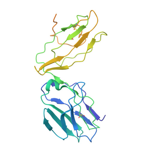
T cell receptor alpha chain (CDR3 to Gly)261Homo sapiensMutation(s): 0 
Entity Groups
Sequence Clusters30% Identity50% Identity70% Identity90% Identity95% Identity100% Identity
Sequence Annotations
Expand
Reference Sequence
Find similar proteins by:|  3D Structure
Entity ID: 3
MoleculeChains  Sequence LengthOrganismDetailsImage
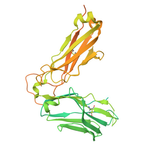
T cell receptor beta chain (CDR3 to Gly)443Homo sapiensMutation(s): 0 
Entity Groups
Sequence Clusters30% Identity50% Identity70% Identity90% Identity95% Identity100% Identity
Sequence Annotations
Expand
Reference Sequence

Experimental Data & Validation

Experimental Data

  • Method: ELECTRON MICROSCOPY
  • Resolution: 3.20 Å
  • Aggregation State: PARTICLE 
  • Reconstruction Method: SINGLE PARTICLE 
EM Software:
TaskSoftware PackageVersion
MODEL REFINEMENTPHENIX1.20.1_4487
RECONSTRUCTIONcryoSPARC

Structure Validation

View Full Validation Report


Entry History 

& Funding Information

Deposition Data


Funding OrganizationLocationGrant Number
Cancer Research UKUnited KingdomCGCATF-2023/100006

Revision History  (Full details and data files)

  • Version 1.0: 2026-11-04
    Type: Initial release