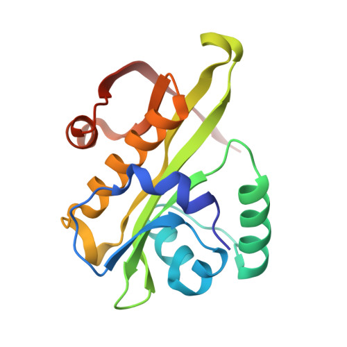
4AG9

C. elegans glucosamine-6-phosphate N-acetyltransferase (GNA1): ternary complex with coenzyme A and GlcNAc


Experimental Data Snapshot

  • Method: X-RAY DIFFRACTION
  • Resolution: 1.76 Å
  • R-Value Free: 0.232 
  • R-Value Work: 0.190 
  • R-Value Observed: 0.191 

Starting Model: experimental
View more details

wwPDB Validation   3D Report Full Report


Ligand Structure Quality Assessment 


This is version 1.3 of the entry. See complete history


Literature

Structural and Biochemical Characterization of a Trapped Coenzyme a Adduct of Caenorhabditis Elegans Glucosamine-6-Phosphate N-Acetyltransferase 1.

Dorfmueller, H.C.Fang, W.Rao, F.V.Blair, D.E.Attrill, H.Van Aalten, D.M.F.

(2012) Acta Crystallogr D Biol Crystallogr 68: 1019

  • DOI: https://doi.org/10.1107/S0907444912019592
  • Primary Citation of Related Structures:  
    4AG7, 4AG9

  • PubMed Abstract: 

    Glucosamine-6-phosphate N-acetyltransferase 1 (GNA1) produces GlcNAc-6-phosphate from GlcN-6-phosphate and acetyl coenzyme A. Early mercury-labelling experiments implicated a conserved cysteine in the reaction mechanism, whereas recent structural data appear to support a mechanism in which this cysteine plays no role. Here, two crystal structures of Caenorhabditis elegans GNA1 are reported, revealing an unusual covalent complex between this cysteine and the coenzyme A product. Mass-spectrometric and reduction studies showed that this inactive covalent complex can be reactivated through reduction, yet mutagenesis of the cysteine supports a previously reported bi-bi mechanism. The data unify the apparently contradictory earlier reports on the role of a cysteine in the GNA1 active site.


  • Organizational Affiliation

    Division of Molecular Microbiology, College of Life Sciences, University of Dundee, Dundee DD1 5EH, Scotland.


Macromolecules
Find similar proteins by:  (by identity cutoff)  |  3D Structure
Entity ID: 1
MoleculeChains Sequence LengthOrganismDetailsImage
GLUCOSAMINE-6-PHOSPHATE N-ACETYLTRANSFERASE
A, B
165Caenorhabditis elegansMutation(s): 0 
EC: 2.3.1.4
UniProt
Find proteins for Q17427 (Caenorhabditis elegans)
Explore Q17427 
Go to UniProtKB:  Q17427
Entity Groups  
Sequence Clusters30% Identity50% Identity70% Identity90% Identity95% Identity100% Identity
UniProt GroupQ17427
Sequence Annotations
Expand
  • Reference Sequence
Small Molecules
Ligands 3 Unique
IDChains Name / Formula / InChI Key2D Diagram3D Interactions
COA
Query on COA

Download Ideal Coordinates CCD File 
C [auth A],
F [auth B]
COENZYME A
C21 H36 N7 O16 P3 S
RGJOEKWQDUBAIZ-IBOSZNHHSA-N
16G
Query on 16G

Download Ideal Coordinates CCD File 
E [auth A],
G [auth B]
2-acetamido-2-deoxy-6-O-phosphono-alpha-D-glucopyranose
C8 H16 N O9 P
BRGMHAYQAZFZDJ-PVFLNQBWSA-N
EDO
Query on EDO

Download Ideal Coordinates CCD File 
D [auth A],
H [auth B]
1,2-ETHANEDIOL
C2 H6 O2
LYCAIKOWRPUZTN-UHFFFAOYSA-N
Experimental Data & Validation

Experimental Data

  • Method: X-RAY DIFFRACTION
  • Resolution: 1.76 Å
  • R-Value Free: 0.232 
  • R-Value Work: 0.190 
  • R-Value Observed: 0.191 
  • Space Group: P 21 21 21
Unit Cell:
Length ( Å )Angle ( ˚ )
a = 48.872α = 90
b = 53.353β = 90
c = 135.096γ = 90
Software Package:
Software NamePurpose
REFMACrefinement
DENZOdata reduction
SCALEPACKdata scaling
MOLREPphasing

Structure Validation

View Full Validation Report


Ligand Structure Quality Assessment 


Entry History 

Deposition Data

Revision History  (Full details and data files)

  • Version 1.0: 2012-07-25
    Type: Initial release
  • Version 1.1: 2012-08-15
    Changes: Database references
  • Version 1.2: 2020-07-29
    Type: Remediation
    Reason: Carbohydrate remediation
    Changes: Data collection, Derived calculations, Other, Structure summary
  • Version 1.3: 2023-12-20
    Changes: Data collection, Database references, Refinement description, Structure summary